
What each examination involves, how long it takes, and what to check in advance.
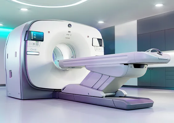
PET-CT is a cutting-edge diagnostic device that combines positron emission tomography (PET) and computed tomography (CT). By injecting fluorodeoxyglucose (FDG), a glucose metabolism indicator, PET visualizes the spread of tumors, metastasis and treatment response. Simultaneous CT imaging pinpoints the location of lesions, and almost the entire body can be evaluated in a scan of about 10 minutes.
Breast PET is a breast-specific PET device capable of detecting small breast cancers. You lie face down with the breast placed in the detector, so no compression is needed. It is performed together with a whole-body PET-CT scan, with no additional FDG injection.
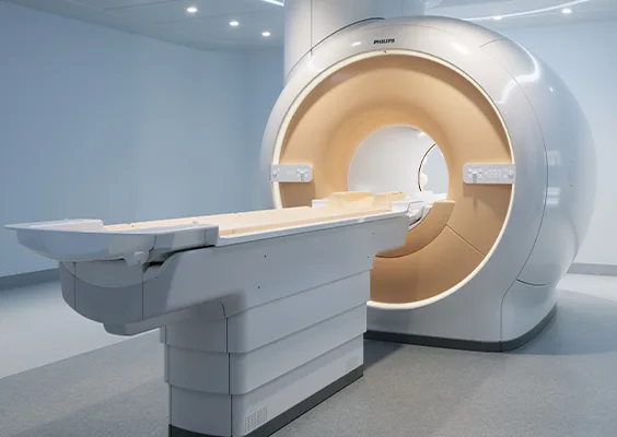
MRI uses a strong magnetic field and radio waves to produce cross-sectional images of the body from various angles, providing detailed information on the brain, spinal cord, internal organs, joints and blood vessels, with no radiation exposure.
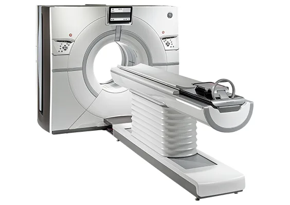
CT irradiates X-rays from around the body and processes them by computer to produce cross-sectional images, revealing internal structures that cannot be seen on regular X-rays. We make every effort to keep radiation exposure to a minimum.
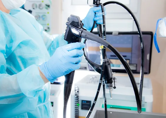
Upper endoscopy uses an endoscope to observe the esophagus, stomach and duodenum. Nasal endoscopy is available on request, please inform us in advance. Those with narrow nasal passages may need oral insertion instead.
You may undergo the examination with sedatives to alleviate anxiety and discomfort (an additional fee applies).
If you cannot tolerate the procedure, no refund is given. Same-day cancellation of the endoscopy cannot be rescheduled.
Slots for PET-CT, MRI and endoscopy are limited. We recommend booking about two months before your preferred date.
Book an appointment →